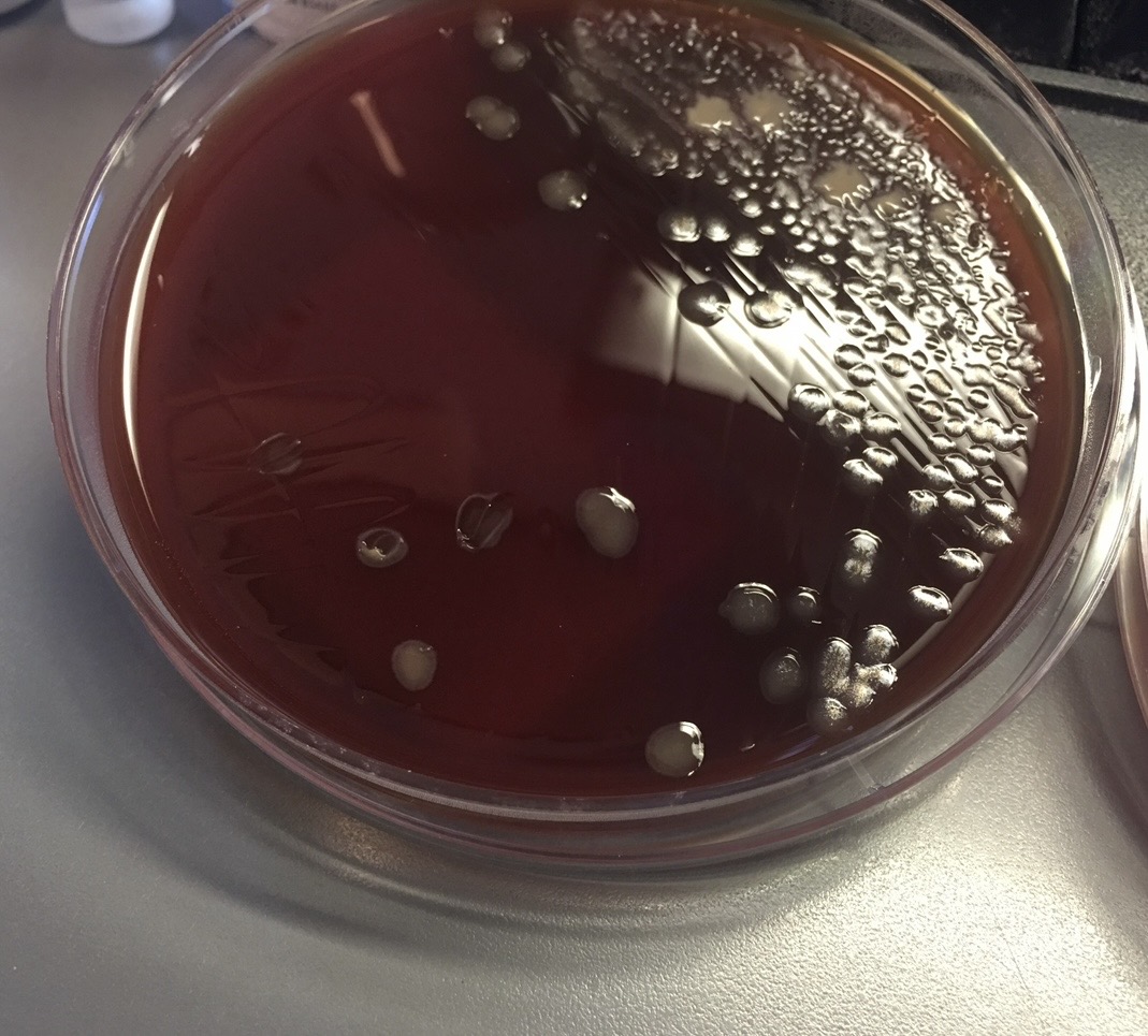
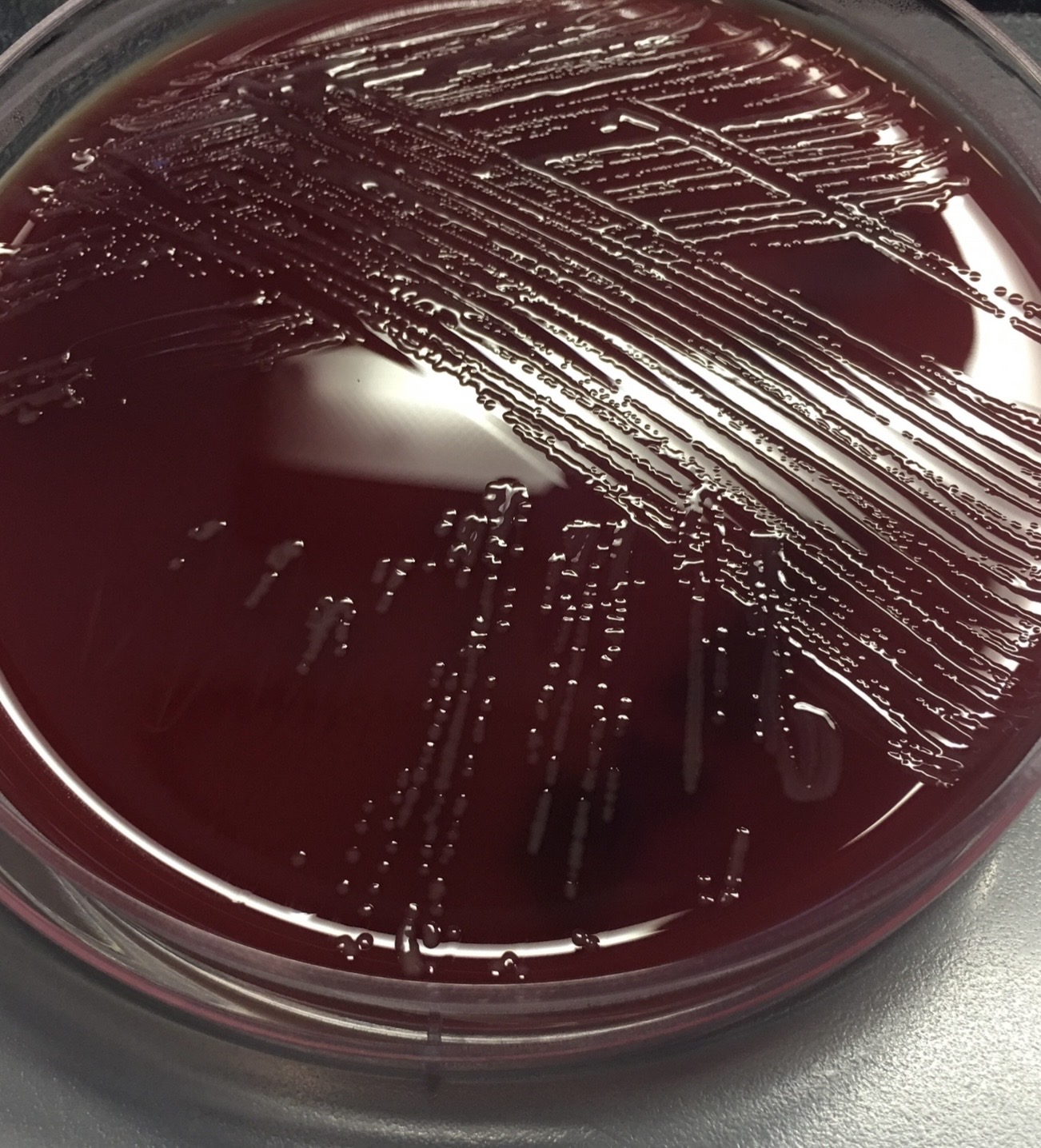
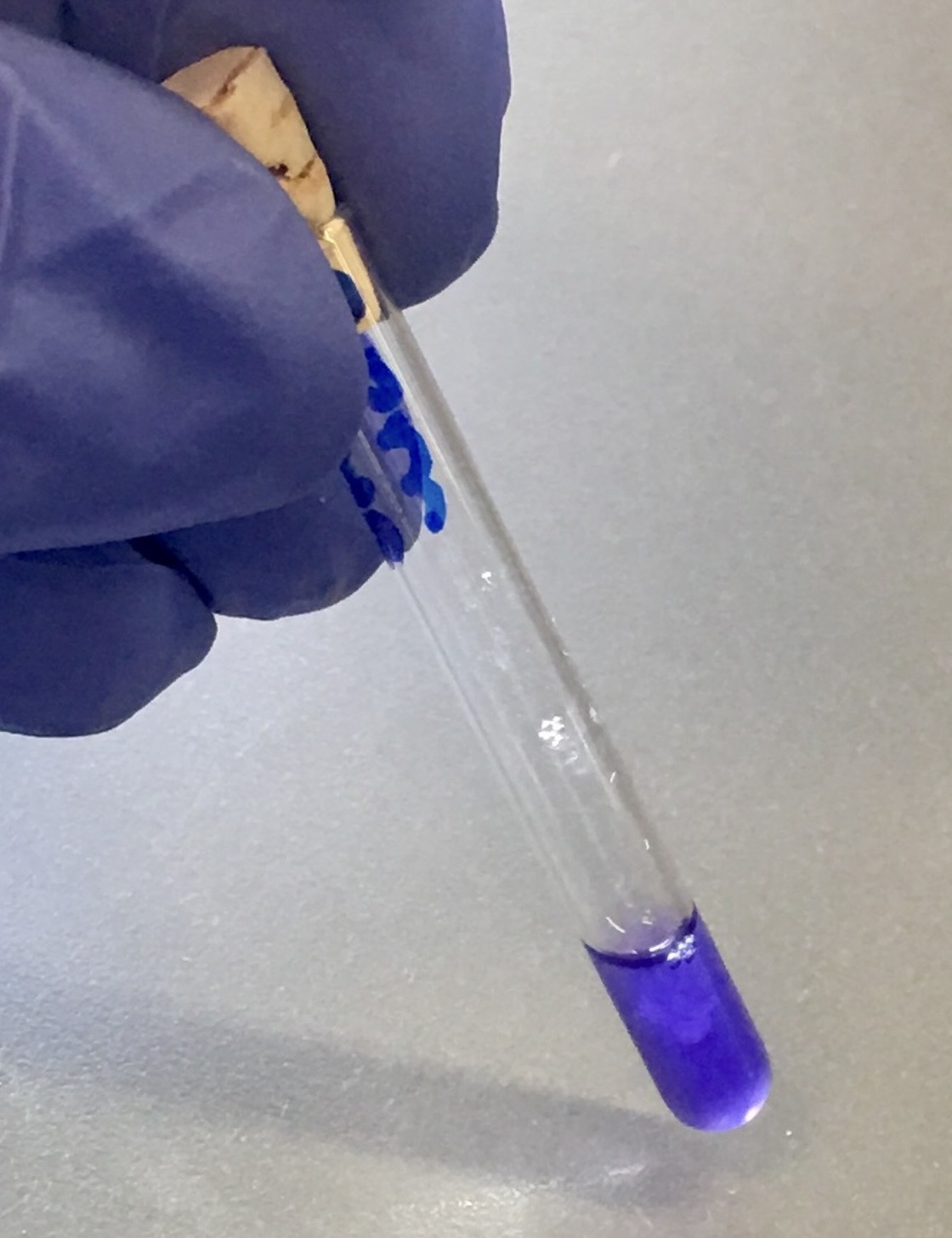
Clinical microbiology, a practical approach to reading the bench

MicroCompass is an educational resource for clinical microbiologists and students. We provide practical laboratory techniques for identification at the bench level.

Use MicroCompass to quickly identify microorganisms, learn microbiology laboratory methods, and assist in bench level decision making.
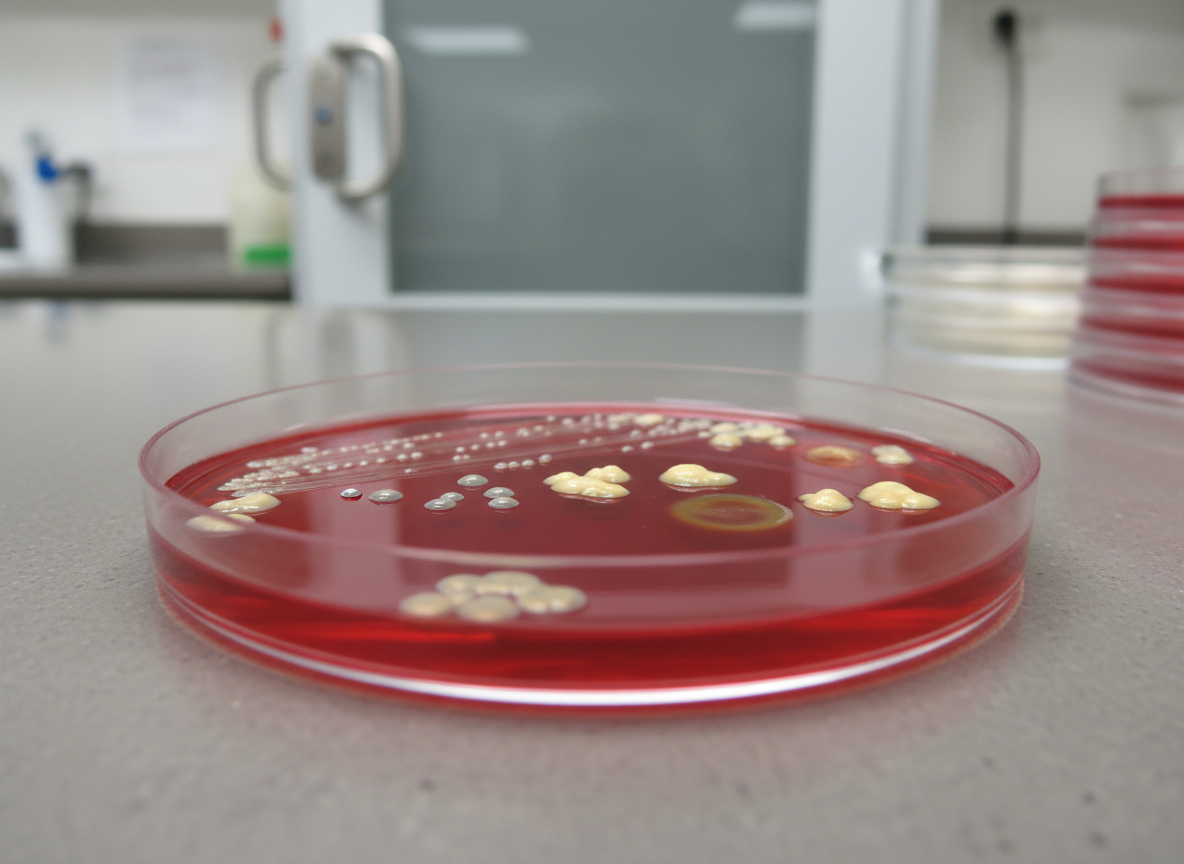
A close-up, photographic realism image of a single streaked blood agar plate, its translucent red surface showing beautifully isolated colonies of different morphologies: small, pinpoint gray colonies, larger creamy ones, and zones of complete and partial hemolysis radiating into the medium. The Petri dish sits on a smooth, neutral gray lab counter, with a blurred background hinting at an incubator door and stacked plates. Cool, even overhead laboratory lighting emphasizes subtle surface gloss and colony texture while casting soft, minimal shadows. Captured at a slight angle with shallow depth of field, the plate occupies the central third of the frame, creating a focused, professional mood ideal for explaining plate reading and colony characteristics.

What’s the Scope?

Latest news and microbiology insights.
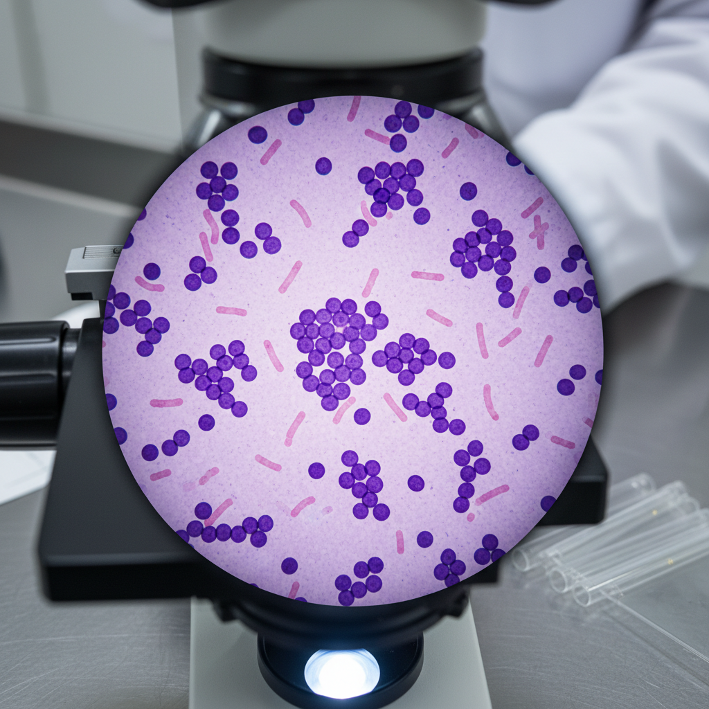
A meticulously detailed, high-magnification photographic-style image of a Gram-stained slide under a clinical light microscope, showing a crisp field of view with deep-violet Gram-positive cocci in clusters and pale pink Gram-negative rods scattered between. The circular microscope field is framed by the dark, slightly out-of-focus rim of the objective lens, suggesting the observer’s perspective. Surrounding the microscope body are subtle hints of a clean lab bench and a rack of unstained slides, rendered as soft bokeh. Bright, cool, coaxial illumination from below produces high contrast and fine detail without glare. The mood is precise and analytical, emphasizing diagnostic interpretation and technique within a professional, educational clinical microbiology context.

Laboratory Techniques

Browse curated clinical microbiology workflows, from specimen collection and culture setup to identification, susceptibility testing, and reporting—each guide emphasizes quality control, biosafety, and reproducible, lab-ready procedures.

Organism Spotlight

A high-resolution, overhead photographic view of a stainless-steel clinical microbiology bench, featuring a neatly arranged array of blood agar, MacConkey, and chocolate agar plates with distinct, well-isolated bacterial colonies in varied colors and textures. Nearby, a calibrated micropipette rests beside a box of sterile tips and a labeled rack of capped culture tubes. The bench surface is spotless and reflective, with a subtle, diffused reflection of the plates. Cool, even fluorescent laboratory lighting illuminates the scene, creating crisp, clinical clarity without harsh shadows. The composition follows a clean, modern aesthetic with strong horizontal lines and generous negative space, evoking precision, order, and professional rigor suitable for an educational microbiology website hero image.

Add your microorganism information here. Include key details such as morphology, staining characteristics, clinical significance, and identification methods.

Contact MicroCompass

Reach out with questions, corrections, or collaboration ideas on clinical microbiology content.

← Back

Thank you for your response. ✨